Abstract
The Emitter Cell produces and releases a small molecule into its surrounding environment, as a means to demonstrate bioproduction within synthetic cells, and small molecule release as a reporter and a means of cell–cell signaling.
Overview
The Emitter Cell produces and releases a chemical signal molecule into the environment. This capacity provides an example of enzymatic small-molecule production, molecule release as a reporter output, inter-cell communication, and co-culture of synthetic cells with living bacteria. The Emitter Cell is largely based on a paper by Jefferson M Smith, Denis Hartmann, and Michael J. Booth: Engineering cellular communication between light-activated synthetic cells and bacteria.
In this first Emitter, the cell produces and releases N-isovaleryl-L-homoserine lactone (IV-HSL). IV-HSL is a branched acyl-homoserine lactone with several advantages for use in the Emitter Cell: it is able to cross the synthetic cell membrane; its uncommon branched-chain structure makes it orthogonal from many other HSLs Lindemann, 2011; and it is able to activate expression in signal receiver cells (in this case, E. coli) at very low (picomolar) concentrations Lindemann, 2011. The IV-HSL signal is received by a population of E. coli cells, which respond by producing a fluorescent output.
The Detector Cells and Emitter together form the basis for an upcoming Responder Cell; a synthetic cell which can detect a molecular input (such as aTc or IV-HSL itself), and produce a molecular output (IV-HSL) in response. Coupling Detector and Emitter modules will enable signal amplification, where a low amount of a molecule of interest can activate a large population of Responder cells and generate an output that is easy to detect. IV-HSL-detecting Responder Cells could also detect the production of IV-HSL from living cells, providing a means to report on their state in co-culture.
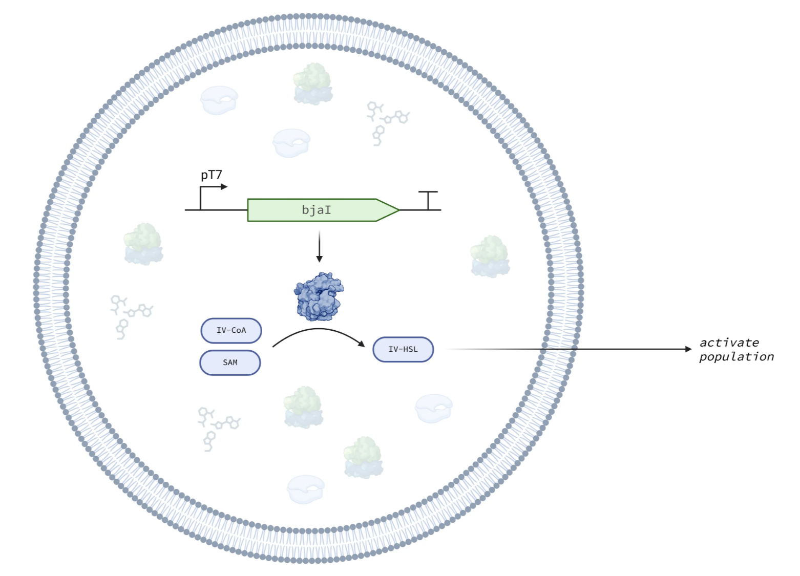
Figure 1:Schematic of IV-HSL emitter cell.
Design
The Emitter Cell implements the IV-HSL Emitter Module within a synthetic cell. The Emitter Module produces the BjaI enzyme under the control of a constitutive T7 promoter. BjaI produces IV-HSL from two substrate molecules, S-adenosylmethionine (SAM) and isovaleryl coenzyme A (IV-CoA). IV-HSL diffuses out of the cell, through the lipid bilayer.
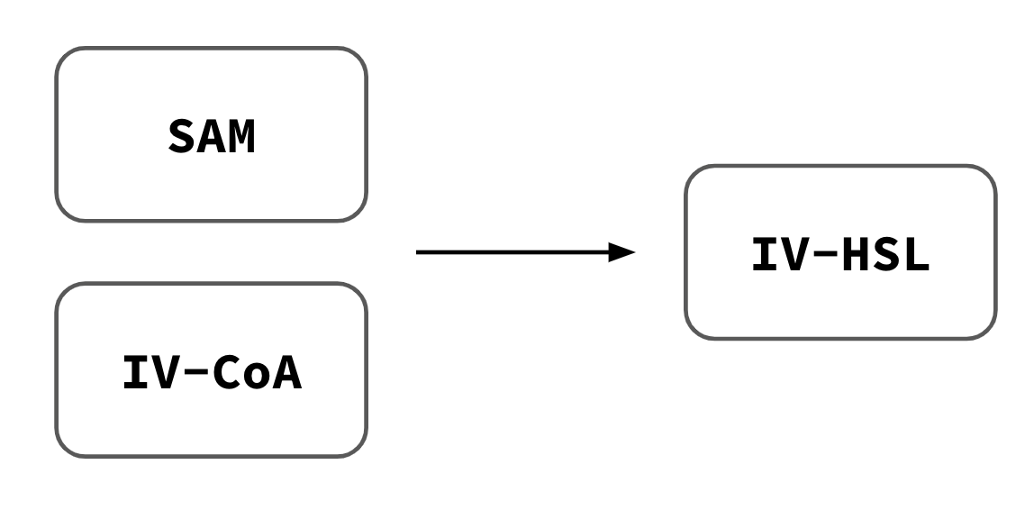
Figure 2:Schematic of IV-HSL emitter module.
Usage
Protocol
Protocol: IV-HSL Emitter Module
Modules
DNA Components
pT7-baI -- Nucleus v0.2.0 Distribution Plate upcoming. Expresses the BjaI enzyme to produce IV-HSL.
bjaR-GFP-native -- Nucleus v0.2.0 Distribution Plate upcoming. E. coli native receiver module; responds to IV-HSL by producing GFP.
Key Materials
| Name | Product | Manufacturer | Part # | Price | Link |
|---|---|---|---|---|---|
| SAM | S-adenosylmethionine (SAM) | NEB | B9003S | $45 | [link] |
| IV-CoA | Isovaleryl coenzyme A lithium salt hydrate | Millipore Sigma | I9381-10MG | $348 | [link] |
| IV-HSL | 3-Methyl-N-[(3S)-tetrahydro-2-oxo-3-furanyl]butanamide | LGC | TRC-M282980-50MG | $171 | [link] |
Performance Data
Emitter Cells were constructed following and co-cultured with E. coli containing the bjaR-GFP-native IV-HSL receiver plasmid. We performed time-series confocal microscopy (Revvity Operetta CLS) over 8 hours, collecting red (Rhodamine-B) and green (GFP) fluorescence, and brightfield images at 40x magnification across multiple fields per well, such that the entirety of each well was imaged. Timepoints were approximately 15 minutes apart.
The Emitter Cell causes E. coli to express GFP in response to IV-HSL.
Emitter Cell Timeseries. (Positive) Liposomes contain PURE and 100 nM IV-HSL. (Negative) Liposomes contain PURE supplemented with SAM and IV-HSL, but no DNA encoding BjaI. (Emitter) Liposomes contain PURE expressing BjaI from pT7-bjaI. Exposures are matched between wells. Each field of view is 167 uM wide.
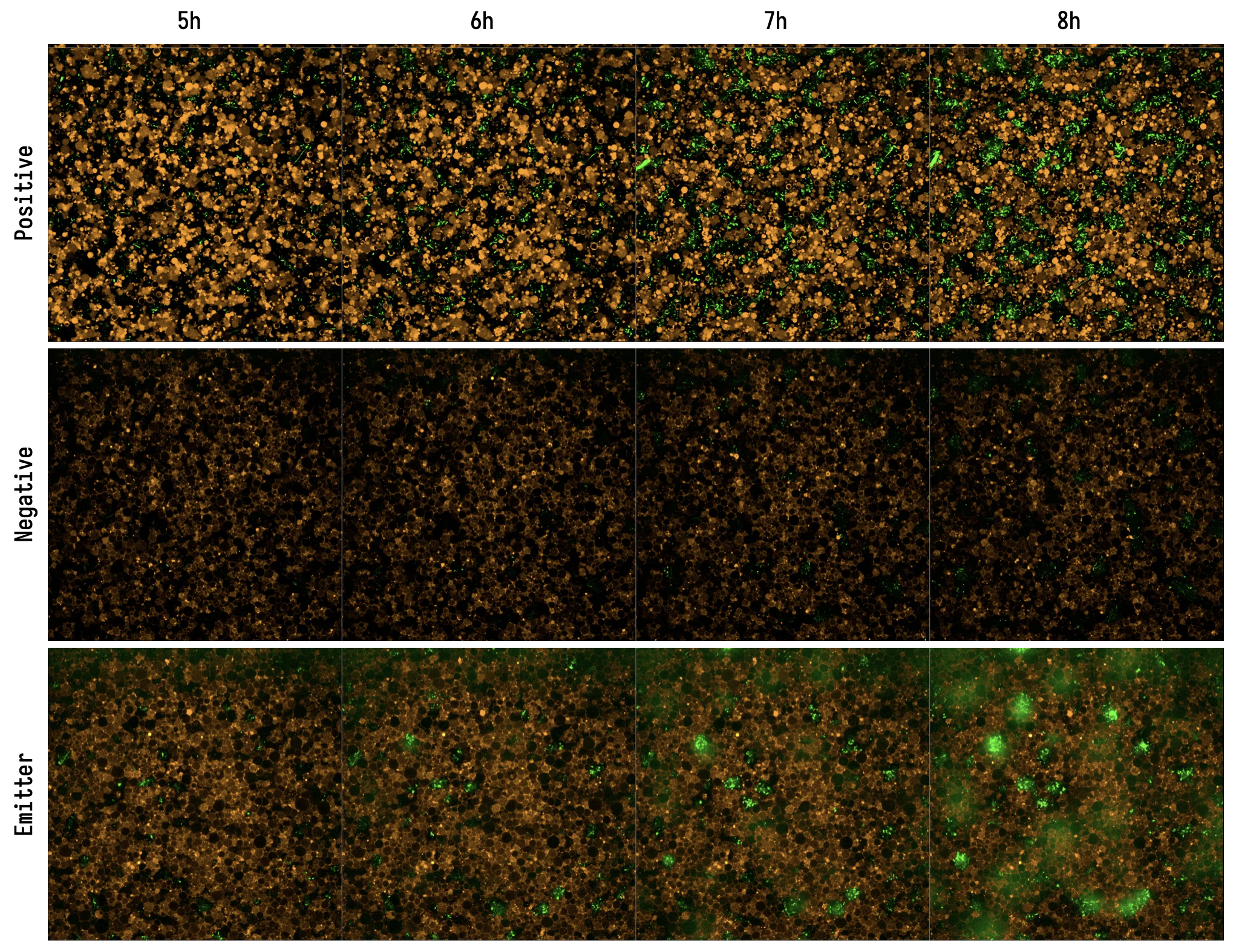
Liposomes exclude E. coli cells from the plate coverslip
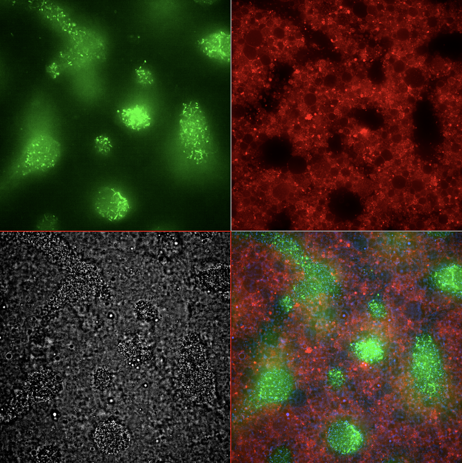
Figure 5:Emitter Cell Endpoint Montage. Single field of view of Emitter Cell in co-culture with E. coli receiver cells at t = 8 hours. (green) E. coli producing GFP in response to IV-HSL emitted by the emitter cells. (red) Emitter cells with rhodamine-labeled membrane producing IV-HSL. (grey) Brightfield image of liposomes and E. coli cells. (rgb) Merged image.
Figure 6:Emitter Cell Endpoint Z-Stack. Z-stack, single field of view of the Emitter Cell after the final timeseries timepoint (>8h). Liposomes preferentially form a layer on the surface of the cover slip, occluding many of the E. coli cells from the bottom layer imaged during the timeseries. More activated E. coli cells become visible at longer focal distances (higher in the liquid column of the well).
Credits
- Jefferson Smith & Michael Booth (Oxford / UCL)
- b.next
- Smith, J. M., Hartmann, D., & Booth, M. J. (2023). Engineering cellular communication between light-activated synthetic cells and bacteria. Nature Chemical Biology, 19(9), 1138–1146. 10.1038/s41589-023-01374-7
- Lindemann, A., Pessi, G., Schaefer, A. L., Mattmann, M. E., Christensen, Q. H., Kessler, A., Hennecke, H., Blackwell, H. E., Greenberg, E. P., & Harwood, C. S. (2011). Isovaleryl-homoserine lactone, an unusual branched-chain quorum-sensing signal from the soybean symbiont Bradyrhizobium japonicum. Proceedings of the National Academy of Sciences, 108(40), 16765–16770. 10.1073/pnas.1114125108